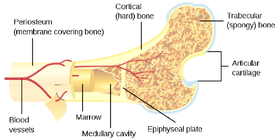
Bone Cross Section Anatomy / Compact Bone Diagram Anatomy Bones Biology Diagrams Human Anatomy Drawing : Spongy bone and compact bone.. Cross section of a bone, this image shows the interior of the bone, which has a lot of spongy bone tissue. Learning cross sectional brain anatomy in this section, you will be presented with several images and figures illustrating the cross sectional brain anatomy that you will be learning. Foot bone anatomy x ray 12 photos of the foot bone anatomy x ray foot bone anatomy x ray, bone, foot bone anatomy x ray. The wider section at each end of the bone is called the epiphysis (plural = epiphyses), which is filled with spongy bone. 12 photos of the cross section of human bone diagram.
As we age, our bones lose their strength. New users enjoy 60% off. Data and dicom images archived on the pacs (picture archiving and communicating system) were processed and exported as jpeg images. A typical long bone shows the gross anatomical characteristics of bone. They are obtained by taking imaginary slices perpendicular to the main axis of organs, vessels, nerves, bones, soft tissue, or even the entire human body.
Cross Section Of The Head Of The Femur Showing Normal Bone Marrow Versus License Download Or Print For 30 32 Photos Picfair from res.cloudinary.com With regards to bone anatomy, some of the parts that can be easily identified include: Two types of bone tissues in cross section of a long bone : Internal structure of a human long bone, with a magnified cross section of the interior. Photomechanical print page item number: The upper (biting) surfaces of the tooth are at top, with the lower sections (bottom) embedded in the gums and jaw bone (not shown). Anatomy of a flat bone. A typical long bone shows the gross anatomical characteristics of bone. They are obtained by taking imaginary slices perpendicular to the main axis of organs, vessels, nerves, bones, soft tissue, or even the entire human body.
As with other tools applied to petroleum development.
The upper (biting) surfaces of the tooth are at top, with the lower sections (bottom) embedded in the gums and jaw bone (not shown). As with other tools applied to petroleum development. 12 photos of the cross section of human bone diagram. Spongy bone and compact bone. The central tubular region of the bone, called the diaphysis, flares outward near the end to form the metaphysis, which contains a largely cancellous, or spongy, interior. Anatomies like brain, temporal bone/internal auditory meatus, nasopharynx, orbit, paranasal sinuses, cranial nerves, temporomandibular joint, neck, brachial plexus, spine, shoulder, arm, elbow, forearm, wrist, hand, finger, thumb, thorax/lung, coronary arteries, abdomen, pelvis, hip, thigh, knee, leg, ankle, foot, angiogram, etc. It is to be hoped that a basic understanding of the anatomy of the temporal bone, and a systematic approach to the various pathologic entities that can affect it, can reduce the anxiety associated with, and improve. Looking at a bone in cross section, there are several distinct layered regions that make up a bone. Bone matrix and cells bone matrix osseous tissue is a connective tissue and like all connective tissues contains relatively few cells and large amounts of extracellular matrix. The wider section at each end of the bone is called the epiphysis (plural = epiphyses), which is filled with spongy bone. It has a unique histological appearance, which enables it to carry out its numerous functions: The outside of a bone is covered in a thin layer of dense irregular connective tissue called the periosteum. Bone on side of the foot
It has a unique histological appearance, which enables it to carry out its numerous functions: It is to be hoped that a basic understanding of the anatomy of the temporal bone, and a systematic approach to the various pathologic entities that can affect it, can reduce the anxiety associated with, and improve. Synovial joint capsule bones chart. The central tubular region of the bone, called the diaphysis, flares outward near the end to form the metaphysis, which contains a largely cancellous, or spongy, interior. As with other tools applied to petroleum development.
9 1 Bone Structure And Function Medicine Libretexts from med.libretexts.org This category is largely categorized by the content of the bone rather than the shape. Download 706 bone cross medical section stock illustrations, vectors & clipart for free or amazingly low rates! As with other tools applied to petroleum development. Data and dicom images archived on the pacs (picture archiving and communicating system) were processed and exported as jpeg images. New users enjoy 60% off. Bone is a specialised type of connective tissue. Internal structure of a human long bone, with a magnified cross section of the interior. Antique illustration of human body anatomy bones, skull:
It has a unique histological appearance, which enables it to carry out its numerous functions:
Two types of bone tissues in cross section of a long bone : Antique illustration of human body anatomy bones, skull: Learning cross sectional brain anatomy in this section, you will be presented with several images and figures illustrating the cross sectional brain anatomy that you will be learning. These canals are part of the osteon structure of the cortex. Ct of the petrous bone an axial and coronal bone multislice computed tomography imaging of the temporal bone was performed on a normal subject. Compact bone is the outer layer and the spongy bone forms the inner layer. Smartdraw includes 1000s of professional healthcare and anatomy chart templates that you can modify and make your own. Download 706 bone cross medical section stock illustrations, vectors & clipart for free or amazingly low rates! Red marrow fills the spaces in the spongy bone. The wider section at each end of the bone is called the epiphysis (plural = epiphyses), which is filled with spongy bone. Browse 4,287 bone cross section stock photos and images available, or search for human bone cross section to find more great stock photos and pictures. Bone on side of the foot Discover (and save!) your own pins on pinterest
For each level or axial section, there will be an actual ct brain image that is labeled and figures depicting where the scan is in the body. As the names suggest compact bone looks compact and the spongy bone looks like sponges. Cross section diagram of human bone, bone, cross section diagram of human bone. Cross section of a bone, this image shows the interior of the bone, which has a lot of spongy bone tissue. New users enjoy 60% off.
Internal Anatomy Of Bone Femur Spc Id 3092 Science 3d Illustration from www.sciencepicture.co It includes such bones as the hip and vertebrae. Antique illustration of human body anatomy bones, skull: Foot bone anatomy x ray 12 photos of the foot bone anatomy x ray foot bone anatomy x ray, bone, foot bone anatomy x ray. Browse 4,287 bone cross section stock photos and images available, or search for human bone cross section to find more great stock photos and pictures. As we age, our bones lose their strength. 12 photos of the cross section of human bone diagram. Two types of bone tissues in cross section of a long bone : Browse 4,275 bone cross section stock photos and images available, or search for human bone cross section to find more great stock photos and pictures.
The central tubular region of the bone, called the diaphysis, flares outward near the end to form the metaphysis, which contains a largely cancellous, or spongy, interior.
It has a unique histological appearance, which enables it to carry out its numerous functions: New users enjoy 60% off. Bone on side of the foot This category is largely categorized by the content of the bone rather than the shape. Smartdraw includes 1000s of professional healthcare and anatomy chart templates that you can modify and make your own. Download 706 bone cross medical section stock illustrations, vectors & clipart for free or amazingly low rates! The compact bone is made up of osteon. Related posts of cross section of a long bone foot bone anatomy x ray. Synovial joint capsule bones chart. Antique illustration of human body anatomy bones, skull: For each level or axial section, there will be an actual ct brain image that is labeled and figures depicting where the scan is in the body. Ct of the petrous bone an axial and coronal bone multislice computed tomography imaging of the temporal bone was performed on a normal subject. Photomechanical print page item number:
It has a unique histological appearance, which enables it to carry out its numerous functions: bone cross section. Foot bone anatomy x ray 12 photos of the foot bone anatomy x ray foot bone anatomy x ray, bone, foot bone anatomy x ray.


0 Komentar