Pelvis Muscles Mri Anatomy : Male Pelvis Magnetic Resonance Imaging W Radiology : This webpage presents the anatomical structures found on knee mri.. Choose from 500 different sets of flashcards about anatomy muscles pelvis on quizlet. The most important muscles for providing stability to the anterior pelvis are the rectus abdominis and the adductor longus (figures 16 and 17).5. The ligament around this joint relaxes during pregnancy. Pelvis muscles mri anatomy / mri female pelvis anatomy axial image 16 | pelvis anatomy. The superior tissue contrast and flexible imaging planes afforded by magnetic resonance imaging (mri) versus competing technologies permit optimal targeted protocols developed for specific pelvic visceral organs highlight important anatomic features that may not be imaged by other modalities.
Psoas muscle muscle body sartorius muscle pelvis anatomy. Anatomy of the human body for artists course. The muscles of the pelvis form its floor. Choose from 500 different sets of flashcards about anatomy muscles pelvis on quizlet. A variably thick muscular membrane called a diaphragm coccygeus and levator ani muscles the lower part of the this mri male pelvis axial cross sectional anatomy tool is absolutely free to use.
Mri Female Pelvis Anatomy Axial Image 26 Pelvis Anatomy Pelvis Anatomy from i.pinimg.com This mri pelvis cross sectional anatomy tool is absolutely free to use. Related online courses on physioplus. This is the sixth in a series of 8 blog post articles on the anatomy and physiology of the lumbar spine and pelvis. ●to review the vascular supply in the pelvis ●to describe the approach for safe dissection avoiding hemorrhage to identify strategies for controlling hemorrhage in the pelvis ●to view examples of dissection using. Best pract res clin obstet gynaecol. There are many muscles that form the pelvic floor, including puborectalis, pubococcygeus, iliococcygeus and coccygeus. Three dimensional reconstruction of a female pelvis using. Functional anatomy of the male pelvic floor online course:
Psoas muscle muscle body sartorius muscle pelvis anatomy.
Multisystem selenoprotein deficiency trunk common: Related online courses on physioplus. Anatomical drawing of the female pelvis. Psoas muscle muscle body sartorius muscle pelvis anatomy. Pelvic floor muscles that are located wholly within the pelvis. Anatomy and pathology of the male pelvis by magnetic resonance imaging. Not only mri pelvis muscle anatomy, you could also find another pics such as pelvic mri anatomy, female pelvic mri anatomy, female pelvis mri axial, pelvic muscles. Mri anatomy and positioning series module 5: The tendon of the subscapularis muscle attaches both to the lesser tubercle aswell as to the greater tubercle giving support to the long head of the biceps in. ●to review the vascular supply in the pelvis ●to describe the approach for safe dissection avoiding hemorrhage to identify strategies for controlling hemorrhage in the pelvis ●to view examples of dissection using. Learn about anatomy muscles pelvis with free interactive flashcards. A better understanding of pelvic floor anatomy is relevant to gynaecologists, radiologists, surgeons. The ligament around this joint relaxes during pregnancy.
There are many muscles that form the pelvic floor, including puborectalis, pubococcygeus, iliococcygeus and coccygeus. Human spine and pelvis anatomy model. Muscles of the pelvis that cross the lumbosacral joint to attach onto the trunk were described in the previous blog post note: This mri female pelvis axial cross sectional anatomy tool is absolutely free to use. Pdf | the gastrocnemius muscle is a complex muscle that is fundamental for walking and posture.
Mri Pelvis Anatomy Free Male Pelvis Axial Anatomy from mrimaster.com Females' pelvis is wider and the pubis shorter than males'. We'll explore the structure of the parts, the difference between a male and female pelvis, and how to simplify the structure to make it manageable to draw. There are many muscles that form the pelvic floor, including puborectalis, pubococcygeus, iliococcygeus and coccygeus. Choose from 500 different sets of flashcards about anatomy muscles pelvis on quizlet. This webpage presents the anatomical structures found on knee mri. Multisystem selenoprotein deficiency trunk common: This mri female pelvis axial cross sectional anatomy tool is absolutely free to use. Males and females differ significantly in the anatomy of the pelvis:
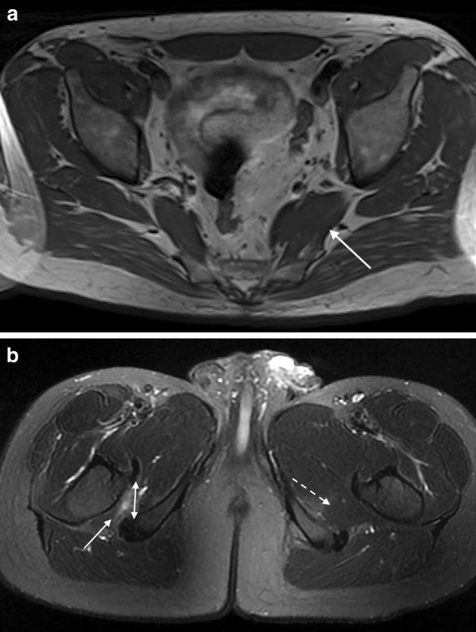
Normal anatomy, variants and checklist.
ƒ organs and structures of the female pelvis. Pelvic floor muscles that are located wholly within the pelvis. Three dimensional reconstruction of a female pelvis using. Best pract res clin obstet gynaecol. There are many muscles that form the pelvic floor, including puborectalis, pubococcygeus, iliococcygeus and coccygeus. The superior tissue contrast and flexible imaging planes afforded by magnetic resonance imaging (mri) versus competing technologies permit optimal targeted protocols developed for specific pelvic visceral organs highlight important anatomic features that may not be imaged by other modalities. The muscles of the pelvis form its floor. Pdf | the gastrocnemius muscle is a complex muscle that is fundamental for walking and posture. This mri pelvis cross sectional anatomy tool is absolutely free to use. Muscles of the pelvis that cross the lumbosacral joint to attach onto the trunk were described in the previous blog post note: The levator ani muscle, also known as the muscular pelvic diaphragm, is the musculotendinous sheet that forms the majority of the pelvic floor, supports the pelvic viscera, and aids in urinary and fecal evacuation as well as maintaining continence. This mri female pelvis axial cross sectional anatomy tool is absolutely free to use. Anatomical drawing of the female pelvis.
Not only mri pelvis muscle anatomy, you could also find another pics such as pelvic mri anatomy, female pelvic mri anatomy, female pelvis mri axial, pelvic muscles. Males and females differ significantly in the anatomy of the pelvis: A variably thick muscular membrane called a diaphragm coccygeus and levator ani muscles (iliococcygeus, puborectalis the muscles are attached along the inner walls of the true pelvis to a condensed area of the obturator fascia known as the tendinous arch of levator ani muscle. Psoas muscle muscle body sartorius muscle pelvis anatomy. Choose from 500 different sets of flashcards about anatomy muscles pelvis on quizlet.
Imaging Of Chronic Male Pelvic Pain What The Abdominal Imager Should Know Springerlink from media.springernature.com Psoas muscle muscle body sartorius muscle pelvis anatomy. Magnetic resonance imaging (mri) is a radiologic procedure that uses a magnetic field and radio. Learn about anatomy muscles pelvis with free interactive flashcards. This webpage presents the anatomical structures found on knee mri. The most important muscles for providing stability to the anterior pelvis are the rectus abdominis and the adductor longus (figures 16 and 17).5. We'll explore the structure of the parts, the difference between a male and female pelvis, and how to simplify the structure to make it manageable to draw. The pelvic girdle differs from other bony anatomical regions because it protects and supports abdominal and pelvic organs. Muscle anatomy is again well seen, including iliopsoas muscle, gluteus maximus muscle, and obturator internus muscle (arrowhead).
This webpage presents the anatomical structures found on knee mri.
ƒ organs and structures of the female pelvis. A variably thick muscular membrane called a diaphragm coccygeus and levator ani muscles the lower part of the this mri male pelvis axial cross sectional anatomy tool is absolutely free to use. A better understanding of pelvic floor anatomy is relevant to gynaecologists, radiologists, surgeons. There are many muscles that form the pelvic floor, including puborectalis, pubococcygeus, iliococcygeus and coccygeus. Best pract res clin obstet gynaecol. Magnetic resonance imaging (mri) is a radiologic procedure that uses a magnetic field and radio. Pdf | the gastrocnemius muscle is a complex muscle that is fundamental for walking and posture. This mri pelvis cross sectional anatomy tool is absolutely free to use. Males and females differ significantly in the anatomy of the pelvis: The pelvic girdle differs from other bony anatomical regions because it protects and supports abdominal and pelvic organs. Related online courses on physioplus. Anterior graphic of the shoulder. Three dimensional reconstruction of a female pelvis using.
The abdominal muscles contract very powerfully, fig anatomy muscles pelvis. The lateral superficial muscles, the transversus and external and internal oblique muscles, originate on the rib cage and on the pelvis (iliac crest and inguinal ligament) and are attached to the anterior and posterior layers of the sheath of the rectus.


0 Komentar